Magnetic Resonance Imaging

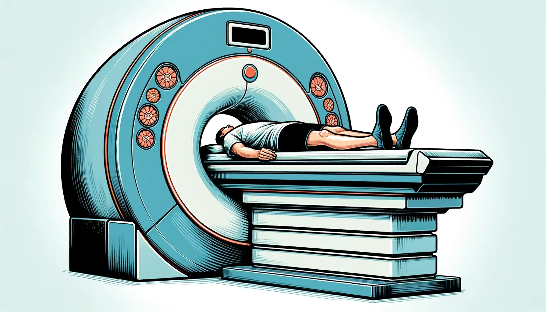
Magnetic Resonance Imaging (MRI) has revolutionized medical diagnostics, offering detailed insights into the human body without the risks associated with ionizing radiation.

Advanced Procedures and Technologies: Biomed Scan Clinic utilizes advanced MRI techniques, such as spectroscopy and specialized protocols for epilepsy, to provide detailed and accurate diagnostics. These procedures offer enhanced imaging capabilities, catering to specific medical needs and conditions.

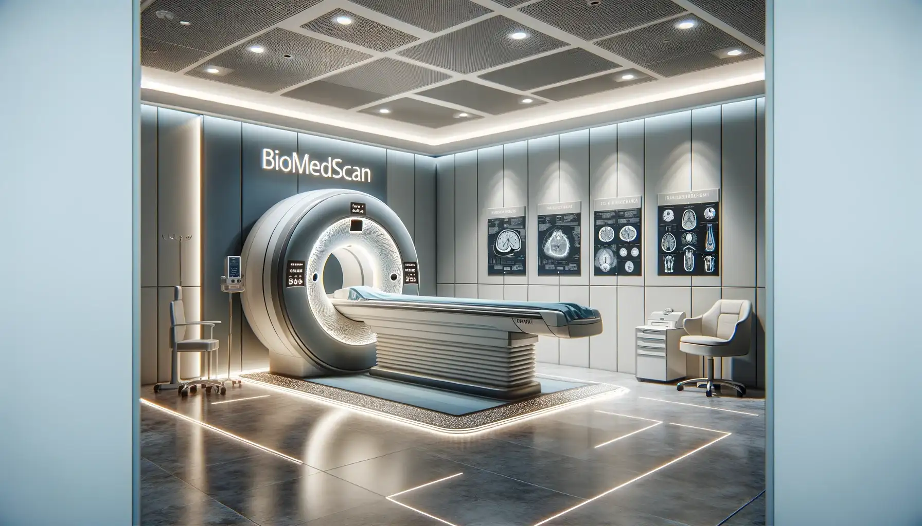
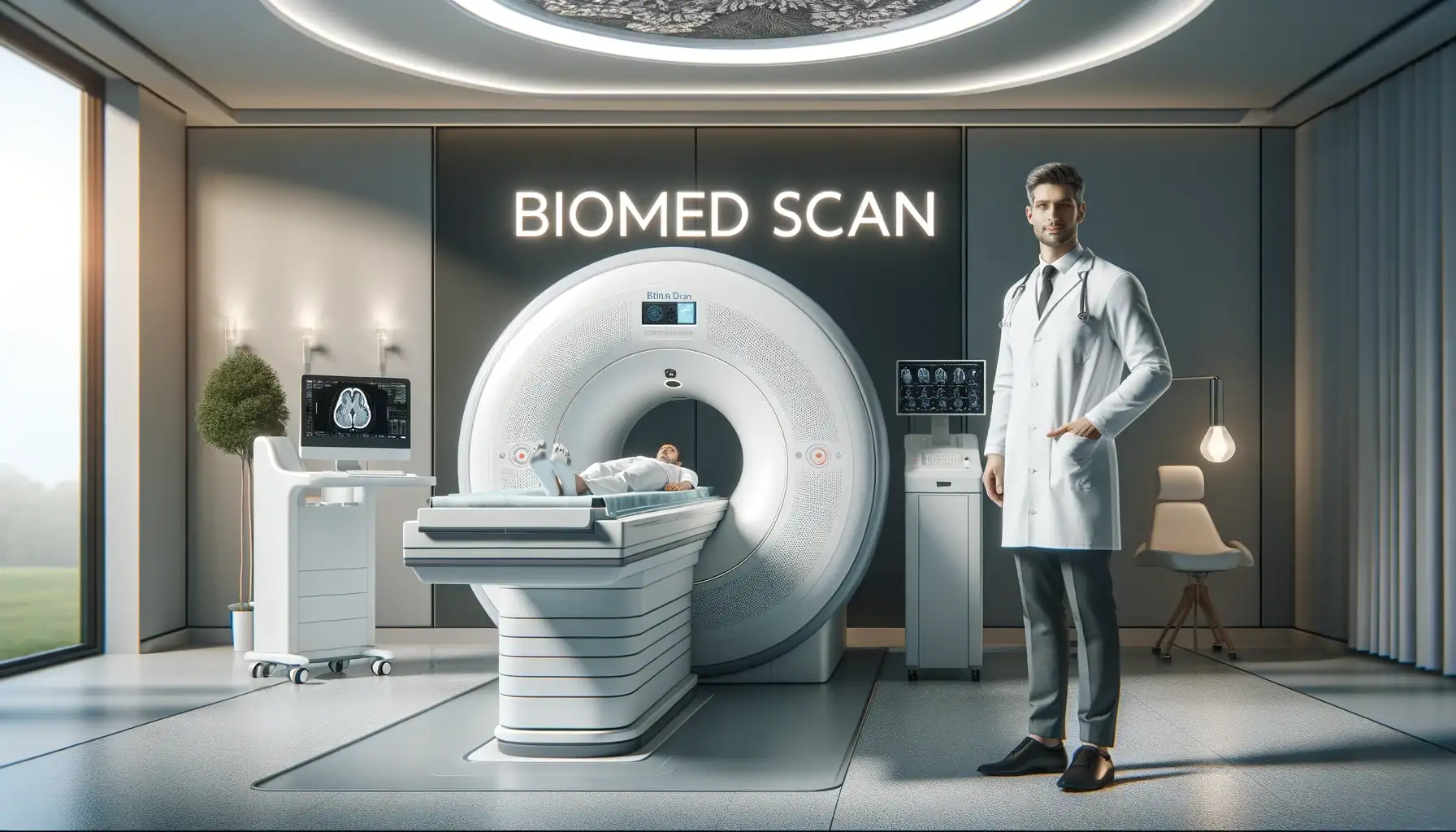
In Bucharest, the forefront of this technological advancement is epitomized by the Biomed Scan Clinic, a center of excellence in MRI services.

Accessibility and Certification

No Ionizing Radiation: MRI is a safer alternative for patients who require multiple scans over time, as it does not expose them to ionizing radiation.

Wide Range of Diagnosable Conditions: MRI at Biomed Scan Clinic is instrumental in diagnosing a diverse array of medical conditions.

Radiology

Medical Imaging

Medical Imaging

Contrast Agent Administration: The contrast agent is only administered to patients who have a recommendation from a specialist and have normal blood tests (creatinine and glomerular filtration rate).

Medical and Technical Aspects of MRI at Biomed Scan Clinic

Pricing and Payment Options: The cost of MRI exams varies depending on the type and complexity of the examination. For instance, a standard Brain MRI costs 590 lei, while specialized procedures like Angio RMN cerebral are priced at 790 lei. Payment options are provided, including the possibility of paying in installments with specific credit cards.

Brain Scan

History and Location

Pregnancy: MRI scans are typically avoided during the first trimester of pregnancy, and pregnant patients should consult their doctor before undergoing an MRI.

Radiology

The clinic’s journey, from its inception to becoming a leading MRI service provider in Bucharest, reflects its dedication to healthcare excellence. With a focus on patient-centric services, Biomed Scan has equipped itself with cutting-edge MRI machines and has cultivated a team of over 24 experienced radiologists and MRI operators, ensuring that each examination is conducted with the utmost precision and care.

Screening for Metal Objects: Due to the strong magnetic field, patients are screened for any metal objects or implants before an MRI scan.

Join us as we navigate through the intricacies of RMN Bucuresti services at Biomed Scan Clinic, unveiling a world where technology and healthcare converge to provide unparalleled diagnostic accuracy and patient care.

Radiology
Magnetic Resonance Imaging

Safety Measures and Protocols:

Advanced MRI Services in Bucharest: A Detailed Look at Biomed Scan Clinic

The Role and Future of Biomed Scan Clinic in Bucharest's Medical Imaging Landscape

Neuroimaging

Looking ahead, the future of MRI technology in Bucharest, with Biomed Scan Clinic at the forefront, appears promising. The non-invasive, radiation-free nature of MRI makes it an increasingly preferred diagnostic tool, particularly beneficial for young patients, pregnant women, and those requiring repeated screenings.

MRI and X-rays/Ultrasound: MRI offers more detailed images compared to X-rays and ultrasound. X-rays are typically used for imaging bones and joints, while ultrasound is used for soft tissue and organ imaging. MRI, however, provides a more comprehensive view of both soft and hard tissues.

MRI Examination Process:

Neuroimaging

Frequently Asked Questions


  • MRI is a medical imaging technique that uses a magnetic field and computer-generated radio waves to create detailed images of the organs and tissues in the body. 
  • It's widely used in hospitals as a diagnostic tool, particularly for imaging the brain, spine, and joints.


  • In a brain scan, MRI uses a powerful magnetic field, radio waves, and a computer to produce detailed pictures of the brain's structure. 
  • It's an essential tool in neuroimaging, helping to diagnose and monitor brain disorders, injuries, and development.


  • T1 and T2 weighted images are types of MRI scans. T1 images are useful for assessing the anatomy of the brain, while T2 images help in identifying fluid and edema. 
  • These imaging techniques offer different views of tissues and are used based on the diagnostic requirement.


  • A contrast agent, often containing gadolinium, is used in some MRI exams to improve the clarity of the images. 
  • It helps to highlight certain structures or abnormalities more clearly, particularly in blood vessels and tumors.


  • A radiographer, or MRI technologist, is responsible for operating the MRI scanner, positioning the patient, and ensuring MRI safety protocols are followed. 
  • They also assist in optimizing image resolution and quality during the radiological examination.


  • Tesla is the unit of measurement for the strength of the magnetic field used in MRI scanners. 
  • Common clinical MRI scanners operate at 1.5 or 3 Tesla (3T MRI Scanner), but high-field MRI scanners can have strengths up to 7 Tesla or more, offering higher image resolution.


MRI sequences are sets of parameters that dictate how the MRI machine will operate to create images. Spin echo and gradient echo are types of sequences that manipulate the protons' behavior in the body, providing different types of image contrast and resolution.


  • Functional MRI (fMRI) measures brain activity by detecting changes in blood flow, often used in cognitive studies and brain mapping. 
  • Diffusion MRI tracks the diffusion of water molecules in tissue, useful in imaging neural pathways and detecting stroke.


  • Yes, certain conditions may prevent a patient from having an MRI, such as the presence of certain types of metal implants, pacemakers, or claustrophobia. 
  • MRI safety guidelines must always be followed to ensure patient safety.


  • Spectroscopy MRI, or Magnetic Resonance Spectroscopy (MRS), is a specialized MRI technique that provides information about the chemical composition of tissues, rather than just their structure. 
  • It's used in body imaging to assess metabolic changes in diseases like cancer or brain disorders.


  • The coil in MRI hardware, often referred to as the MRI coil, plays a crucial role in both sending and receiving radio waves during the scan. 
  • Different types of coils are used depending on the part of the body being imaged. 
  • For example, a head coil is used for brain imaging, while a larger coil may be used for body imaging. 
  • These coils are essential for enhancing image quality and ensuring accurate diagnosis.


  • High-field MRI refers to MRI systems with a higher strength of the magnetic field, typically above 1.5 Tesla. 
  • These machines, like the 3T MRI scanner, offer better image resolution and shorter scan times. 
  • High-field MRI is particularly beneficial in detailed studies, such as brain or musculoskeletal imaging, where precise anatomical details are crucial.


  • MRI sequences are pre-set protocols that determine how the MRI machine will interact with the magnetic field and radio waves to produce images. 
  • Each sequence, like Spin Echo or Gradient Echo, is designed to highlight different tissue properties and is chosen based on the specific diagnostic requirement. 
  • Sequences affect factors like image contrast, resolution, and the time it takes to perform the scan. 
  • They are pivotal in tailoring the MRI to the specific clinical question at hand.